Basal Cell Carcinoma (BCC)
Basal cell carcinoma (BCC) is the most common type of skin cancer and originates from the basal cells of the epidermis. It typically appears after the age of 40 and is more common in people with fair skin. BCC is more likely to develop in areas of the body exposed to sunlight, such as the face, scalp and neck. According to studies, 80% of BCCs appear on the face, with the nose being the most common area. Although it is less aggressive than other types of skin cancer (such as melanoma), BCC can cause local tissue destruction if not treated in time.
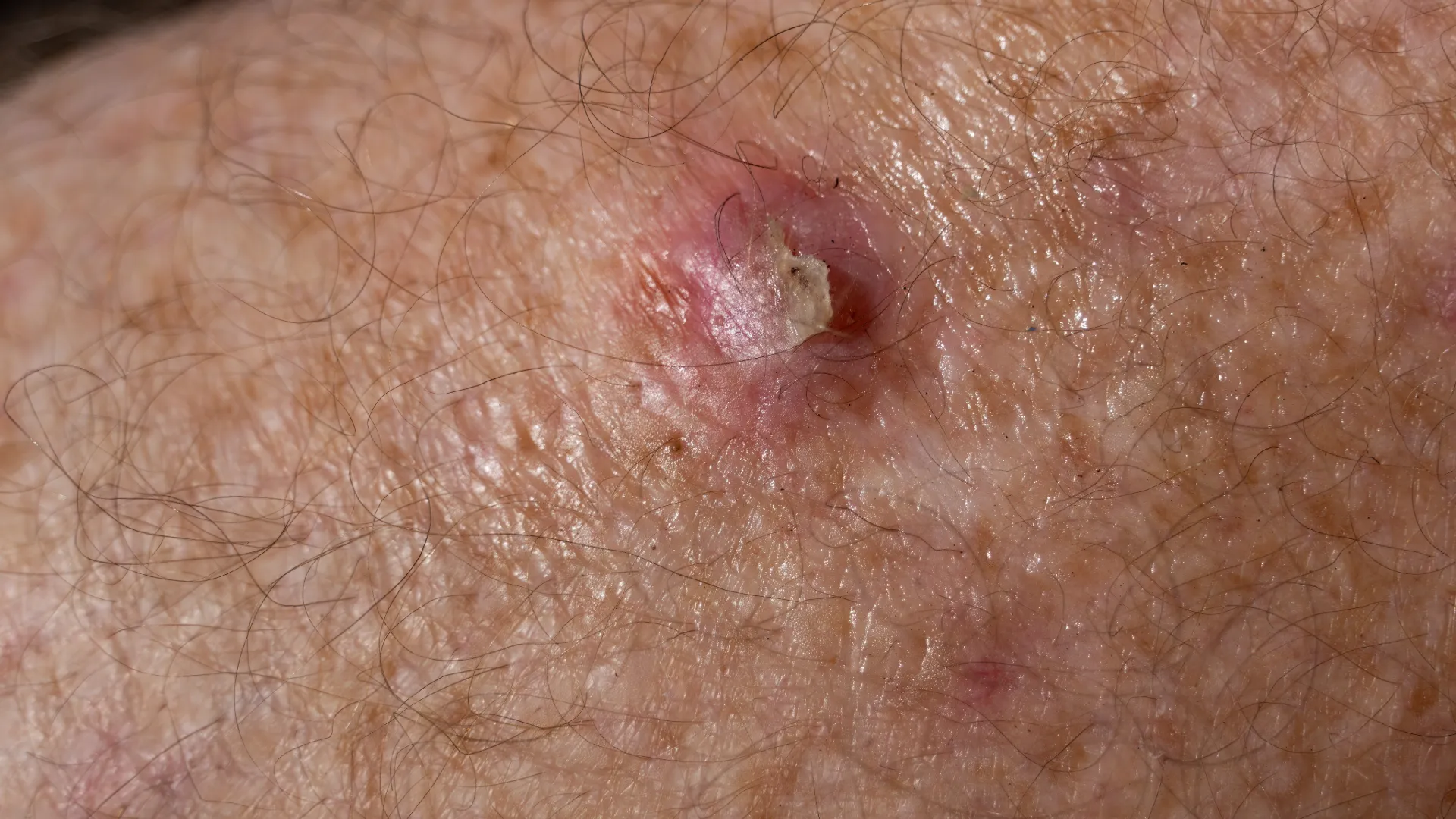
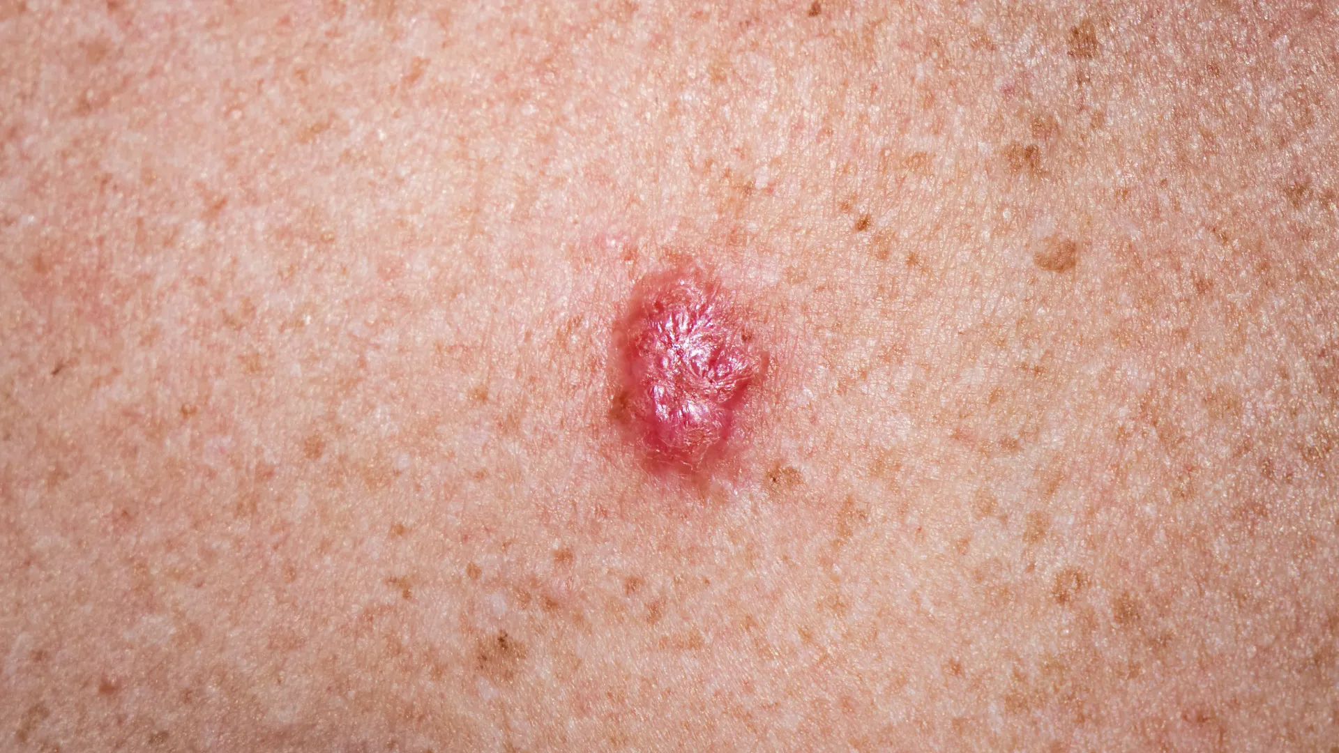
Basal cell carcinoma usually appears as a small, reddish papule or mass that gradually enlarges. Often, the center of the lesion bleeds and may be described by patients as a wound that does not heal for a long period of time. Its development is slow and is usually confined to the skin, without spreading to other parts of the body. Because it has a very low chance of metastasis, the prognosis for BCC is excellent when detected and treated early.
Clinical forms of BCC
BCC is classified into several clinical forms, each with its own characteristics:
Nodular BCC
- Appearance: A white, pearly or pinkish papule that grows slowly and irregularly, forming an oval mass. The center often ulcerates and bleeds.
- Location: Typically found on the face, scalp and hands.
- Aggressiveness: It is the most common and least aggressive form.
Pigmented BCC
- Appearance: A nodular form of BCC that has a dark color due to the presence of melanin. It may be confused with melanoma.
- Location: Most commonly appears on the face or other sun-exposed areas of the body.
- Aggressiveness: Similar to nodular BCC, but more difficult to distinguish due to its pigmented nature.
Superficial BCC
- Appearance: A flat, red or pink plaque with scales, appearing benign, and growing slowly. It is the least aggressive form of BCC.
- Location: Usually found on the trunk or extremities.
- Aggressiveness: Slow development and low aggressiveness, typically not causing damage to surrounding tissues.
Morpheaform BCC
- Appearance: Resembling a scar with ill-defined borders and a yellowish or pale color. It is rarer and more aggressive.
- Location: Often appears on the face and is more difficult to detect due to its uniform color.
- Aggressiveness: Develops deeper and can cause significant local tissue destruction if not treated.
Micronodular BCC
- Appearance: Similar to nodular BCC, but with small islands of cancerous cells extending beyond the visible boundaries of the lesion.
- Location: Commonly appears on areas of the face or parts of the skin with significant sun exposure.
- Aggressiveness: Can grow deeper into the skin and cause more extensive damage.
Prognosis
The prognosis for BCC is excellent, as the likelihood of metastasis is nearly zero. However, if left untreated, BCC can cause local tissue destruction, infiltrating deeper layers of the skin and underlying tissues. The goal of treatment is to completely remove the cancer and restore the anatomical functionality of the affected area.
Treatment
The treatment of BCC depends on its form, location, size and the general health of the patient. The most common treatment methods include:
Surgical Excision
Complete removal of the tumor with a margin of healthy tissue, followed by biopsy.
Cryosurgery
Liquid nitrogen is used to destroy the cancerous tissue. It is ideal for small and superficial lesions.
Mohs Surgery (Mohs Micrographic Surgery)
The most specialized method of surgical removal, particularly useful for tumors located in sensitive areas, such as the face.
Topical Therapy
The application of immunomodulatory creams, such as Imiquimod or 5-Fluorouracil, to treat superficial BCCs.
Radiotherapy
Primarily used for lesions that are not suitable for surgical removal or in cases where other treatments have failed.
Systemic Therapy
Oral medications may be indicated in cases of advanced or metastatic BCC, as well as for patients with multiple BCCs.
Conclusion
Basal Cell Carcinoma (BCC) is one of the most common malignant skin tumors and has an excellent prognosis if diagnosed and treated early. Despite the excellent prognosis, regular monitoring and treatment are required to prevent recurrence or local tissue destruction.