Nevi
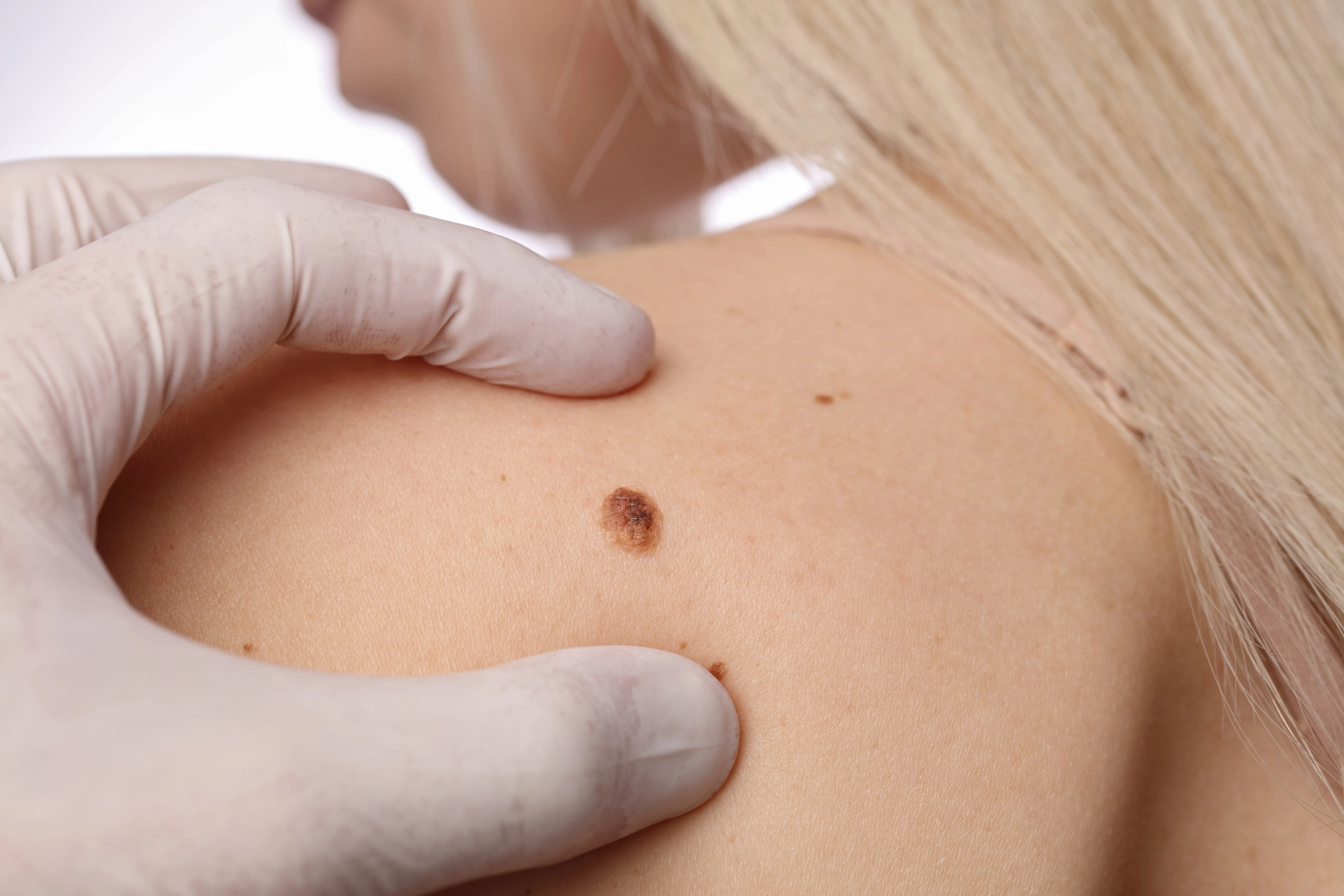
Nevi, also known as moles, are common skin lesions that can appear from infancy. They come in various shapes and colors and can develop anywhere on the body. Nearly all adults have 10-40 nevi.
Depending on when they appear, nevi are classified as congenital (present at birth or develop during infancy) and acquired (develop during childhood or the early years of adulthood). Nevi that develop after the age of 40 should always be checked by a dermatologist.
Types of nevi
Common nevi
These can be congenital or acquired, and are smooth, round with a uniform color (pink, black or brown). They may be flat or slightly raised.
Dysplastic nevi
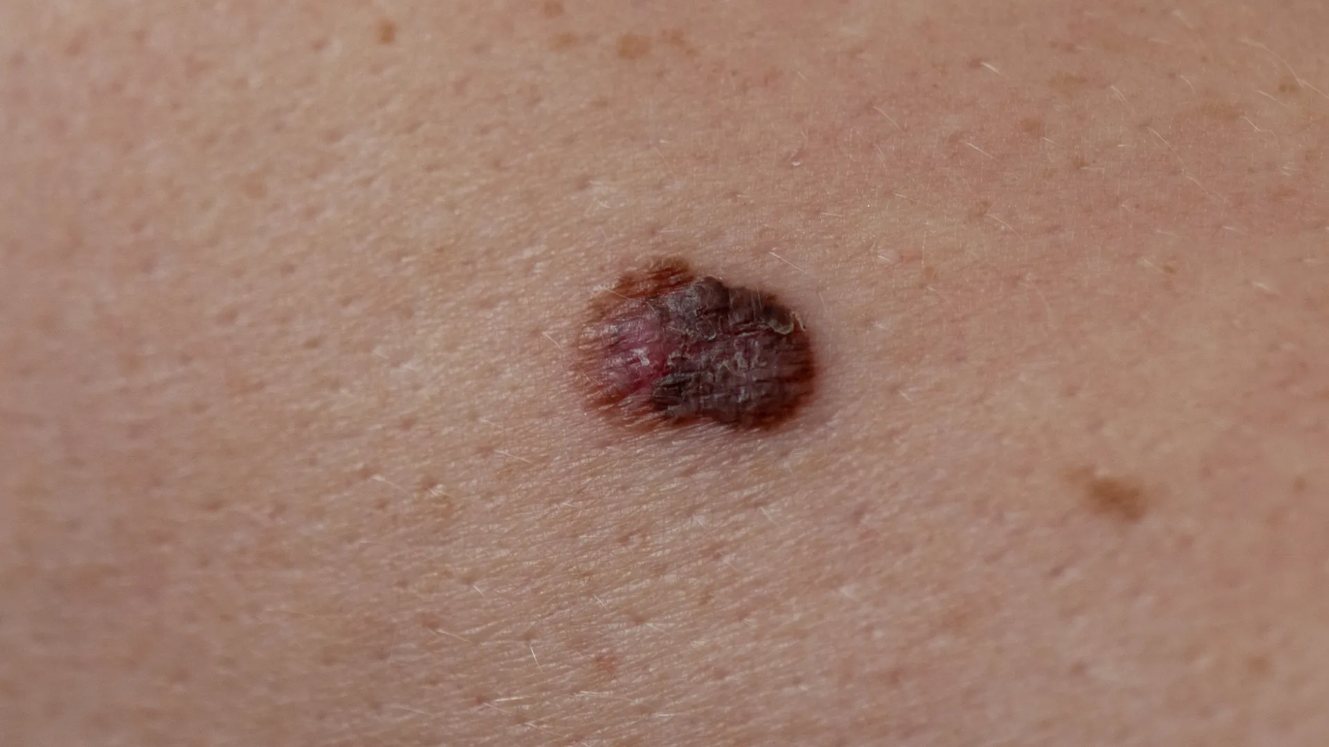
Also called atypical nevi, these are benign (non-cancerous) but may have varying colors, asymmetrical shapes or irregular borders. They are a risk factor for developing melanoma.
Blue nevus
These are dark blue or black nevi, which may be flat or raised. They typically do not change over time.
Sutton’s nevus
This type has a white halo around it. Over time, the color of the nevus fades from the center and may disappear completely. It can coexist with vitiligo (loss of skin pigment).
Becker’s nevus
A pigmented spot that gradually increases in size and may have dense hair on its surface. It can appear anywhere on the body but is most commonly found on the arms or upper trunk.
Ota’s and Ito’s nevi
These are extensive, unilateral pigmented lesions with a characteristic blue tint. These nevi are particularly common in Asian populations. Ota’s nevi usually appear on the face and around the eyes, while Ito’s nevi develop on the upper, lateral chest.
Spitz nevus
A benign lesion typically seen in children and adolescents. It is often a solitary, hemispherical nodule or papule with a pinkish color, usually appearing before the age of 20. It grows rapidly and then remains stable. Rarely, it may be pigmented and is then called Reed’s nevus.
Epidermal nevi
These appear as linear or clustered keratotic or wart-like plaques. They are usually congenital or appear in the first years of life. Rarely, they emerge in adolescence or adulthood and are typically found on the scalp and neck.
Sebaceous nevi
Epidermal nevi that often develop on the scalp, rich in sebaceous glands with residual hair follicles. These nevi are best removed in youth, as the skin on the scalp is more elastic.
Jadassohn’s sebaceous nevus
A skin lesion, typically on the face or scalp, that is present at birth. It consists of sebaceous glands, hair follicles, and ectopic apocrine glands. It appears as a slightly raised plaque, yellow, orange or brown in color, with a smooth or velvety surface. Removal is recommended as it may develop secondary neoplastic changes.
When to be concerned
Nevi that develop or change after the age of 30 require attention and monitoring. To recognize suspicious lesions, the ABCDE method can be used:
- A (Asymmetry): If the nevus is not symmetrical.
- B (Borders): Irregular borders.
- C (Color): Multiple shades in one nevus.
- D (Diameter): A diameter larger than 6 millimeters.
- E (Evolution): Any change in size, shape or color.
Risk factors
Intense sun exposure and sunburns, particularly during childhood, increase the likelihood of developing skin cancer. Additionally, people with a family history of melanoma or with fair skin are at greater risk.
To prevent melanoma, daily sunscreen use and wearing protective clothing is recommended, along with avoiding excessive sun exposure and the use of tanning beds.
Nevus removal
Nevi that are considered suspicious should be removed and biopsied. A nevus may also be removed if it is in a location prone to injury or if it causes aesthetic concerns for the patient.
The method of nevus removal depends on the type of nevus, its location and the patient’s medical history. The main removal methods include:
- Surgical removal: This is the preferred method when malignancy is suspected, as the whole nevus is sent for biopsy.
- Shave biopsy: The protruding part of the nevus is removed and sent for biopsy if necessary. The remaining part is removed with electrocautery or CO2 laser.
- Laser removal: This method is only used when the benign nature of the nevus has been confirmed.

